Treatment of a discoloured, previously traumatised vital tooth UR1
A 30-year-old patient, with a negative medical history, comes to attention with the request to replace the previous composite reconstruction performed 10 years earlier following a trauma of UR1.
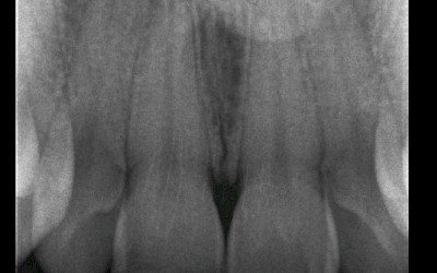
UR1 on clinical examination is responsive to the viability test, and does not present periapical lesions on the radiograph performed on the same day (Fig. 1).
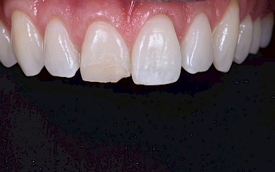
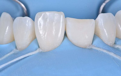
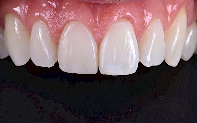
UR1 is discoloured and in a more palatal position than the contralateral central UL1 (Fig. 2).
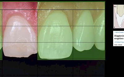
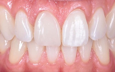
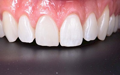
The aesthetic analysis highlights an asymmetry of the gingival zenith between UR1 and UL1. Through the use of a periodontal probe, after plexus anesthesia, the altered passive eruption of the type IA junctional epithelium is confirmed according to the classification of Coslet et al. (Fig. 3).
With a view to carrying out the most conservative restorative treatment possible on the patient, taking into account the age and vitality of the retained dental element, it is decided to carry out a direct composite restoration following planning and a diagnostic wax-up of the case.
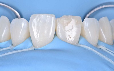
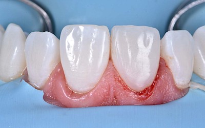
On the day of treatment, following local plexus anesthesia, UR1 is isolated using a rubber dam sheet, extending the isolation to the first premolars (Fig. 4).
Subsequently, the fractured composite reconstruction is removed and a short bevel is performed on the preparation and the entire surface of UR1 is sandblasted with 27μm aluminum oxide powder (Fig. 5).
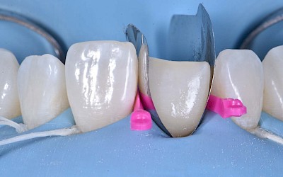
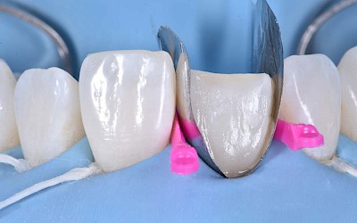
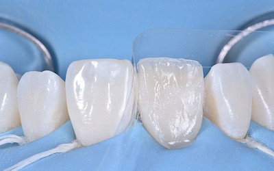
In order to correct the altered passive eruption, it was decided to recreate the emergence profile of the tooth by accentuating the vestibular bulge and seeking symmetry with the contralateral element. For this purpose, a pre-formed metal matrix is used and is blocked with two wedges.
Once the matrix has been adapted, the adhesion procedures are carried out with a 3-step etch&rinse system. Each step is followed by polymerization with UV light for 40 seconds (Fig. 6).
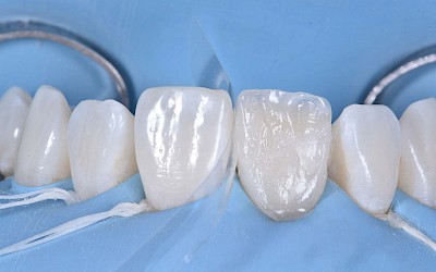
The vestibular emergence profile is recreated with an enamel shade of composite (ESTELITE ASTERIA WE from TOKUYAMA DENTAL) (Fig. 7).
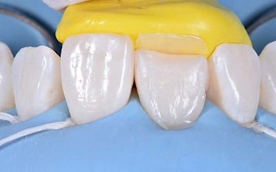
After performing a silicone index of the diagnostic wax-up, the palatine wall is reconstructed by an enamel shade of composite (ESTELITE ASTERIA WE from TOKUYAMA DENTAL) (Fig. 8).
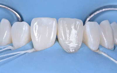
Subsequently, the dentinal anatomy is reconstructed through the reproduction of the mamelons with an opaque dentinal composite shade (ESTELITE SIGMA QUICK OA2 from TOKUYAMA DENTAL); this shade will also be fundamental for correcting the shade of the dischromatic element (Fig. 9).
Light blue and white effect shades (ESTELITE COLOR from TOKUYAMA DENTAL) are applied to emulate the opalescence in the incisal area (Fig. 10).
The layering is completed through the use of an enamel shade (ESTELITE ASTERIA WE from TOKUYAMA DENTAL) in the vestibular with a single addition. The vestibular surface is modelled and controlled in three-dimensional volumes in order to have as few final adjustments as possible. It is then polymerized for 20 seconds and polymerized for 40 seconds in the vestibular and palatine after being covered with glycerin gel to inhibit the hybrid layer of the composite (Fig. 11).
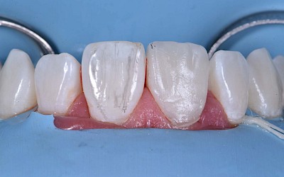
The finishing and polishing procedures are carried out trying to emulate the transition lines of UL1 (Fig. 12 / 13).
The patient is checked again after 21 days (Fig. 14/15) and 12 months (Fig. 16) to evaluate the aesthetic result in shape and colour.

Author:
Dr Nicolò Barbera
Assistant professor in the Division of Cariology and Endodontics at the University of Geneva
Private clinical practice in Lausanne

